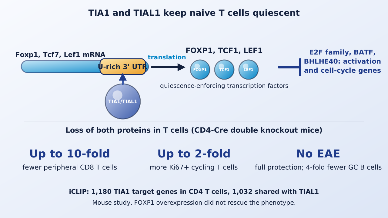
A mouse carries more than a billion naive T cell clones, and most of them sit idle for months or years before meeting an antigen. A French group now reports that two RNA-binding proteins, TIA1 and TIAL1, are needed to hold these cells in that resting state, by making sure that three quiescence-enforcing transcription factors keep being produced.
| Measure | System | Reported result |
|---|---|---|
| Peripheral T cells, double knockout | Spleen and lymph nodes | CD4 down 2 to 5-fold; CD8 down up to 10-fold |
| Germinal center B cells after NP-KLH | Immunized mice | 4-fold fewer |
| EAE after MOG 35-55 | Double knockout mice | Total protection |
| Ki67-positive T cells | Lymph nodes | Up to 2-fold higher |
| Apoptotic T cells | VAD-FMK labeling | 1.5 to 4-fold higher |
| Genes targeted by TIA1 / by both TIA1 and TIAL1 | Activated CD4 T cells, iCLIP | 1,180 / 1,032 |
Background
Quiescent T cells have low metabolic activity and are arrested in G0. Transcription factors such as FOXP1, TCF1 and LEF1 enforce this state, but they have short protein half-lives, so the cells must keep making them. RNA and protein levels correlate poorly in T cells (r² = 0.4), pointing to posttranscriptional control. TIA1 and TIAL1 are paralogs that regulate splicing and translation and act redundantly in B cells, but their role in T cells had not been examined.
Loss of both proteins empties the T cell pool
The authors deleted Tia1 and Tial1 together using CD4-Cre. Thymic T cell development was normal, but peripheral CD4 T cells fell two- to fivefold and CD8 T cells up to tenfold, with more memory- and effector-phenotype cells (more than 10-fold more effector-phenotype CD8 cells). Deleting either gene alone changed nothing, which indicates redundancy. Competitive bone marrow chimeras and tamoxifen-inducible deletion showed that the defect is cell intrinsic. The mice were fully protected from MOG-induced experimental autoimmune encephalomyelitis, and after NP-KLH immunization they had fourfold fewer germinal center B cells and fewer T follicular helper cells.
Cells leave quiescence and die
Up to twofold more T cells were Ki67-positive, BrdU label decayed faster, and activation markers (CD25, CD28, PD-1, CTLA4) were higher on naive and antigen-experienced cells. Apoptotic cells were 1.5- to 4-fold more frequent depending on subset. The defect did not depend on TCR tonic signaling, IL-7–STAT5 signaling, energy metabolism or bulk protein synthesis, and cytokines or TCR stimulation did not rescue survival.
Targets and transcriptome
iCLIP mapped TIA1 and TIAL1 mostly to U-rich elements in introns and 3′ UTRs. In activated CD4 T cells, 1,180 genes were TIA1 targets and 1,032 of them were also TIAL1 targets; TIAL1 targets numbered 3,516 genes in naive and 7,060 in activated CD4 T cells, and 4,053 in naive CD8 T cells. RNA-seq found 1,632 and 2,195 differentially expressed genes in naive and effector-phenotype CD4 T cells, mostly increased, with higher NF-κB, SP1, STAT3, MYC and E2F activity and cell-cycle genes such as Ccnd3, Cdk6 and Pcna. Splicing was also altered: 1,679 events in naive CD4 and 2,022 in naive CD8 T cells, mostly skipped exons with reduced inclusion.
Translation of FOXP1, TCF1 and LEF1
Among 4,094 genes with TIA-bound 3′ UTRs, Foxp1, Tcf7 and Lef1 mRNAs fell 1.5- to 2-fold in knockout cells and their proteins were reduced in naive, memory- and effector-phenotype CD4 and CD8 T cells. In HEK293 reporter assays, wild-type TIA1, but not an RNA-binding mutant lacking RRM1 and RRM2, raised translation from each 3′ UTR without changing mRNA abundance, and knockdown of both proteins lowered it. FOXP1-repressed genes rose and FOXP1-induced genes fell, and E2f1, E2f2, E2f3, E2f7 and E2f8 were higher in knockout cells. Binding to E2f3, Batf and Bhlhe40 3′ UTRs was largely dispensable for their expression.
Keep in mind
All data are from mice and one human cell line used for reporters. The authors report that overexpressing FOXP1 failed to rescue the phenotype, so the effect is multifactorial, and they note that the other targets, including cell-cycle genes, still need single-gene validation. Splicing changes may not be linked to quiescence. The study does not examine human T cells, cancer, or any therapeutic use of TIA1 or TIAL1.
- Paper
- Osma-Garcia IC, Nguyen TMM, Maloudi O, et al. Posttranscriptional regulation by TIA1 and TIAL1 controls the transcriptional program enforcing T cell quiescence. Science Advances. 2026;12(47):eaeh4461. Published November 18, 2026.
- Type
- Mouse genetics, iCLIP and RNA-seq study
- DOI
- 10.1126/sciadv.aeh4461
- PubMed
- PMID 43050523
- Funding
- Agence Nationale de la Recherche (ANR-20-CE15-0007, ANR-22-CE15-0013, ANR-24-CE15-5291); Fondation pour la Recherche Médicale; La Ligue Contre Le Cancer; Cancéropôle Grand Sud-Ouest; ATIP Avenir Plan Cancer. The authors declare no competing interests.
Related articles
 Daily brief · Nov 17, 2026A GCC-Rich Motif Boosts Translation From Non-AUG Start Codons in Human Cells, While eIF1 and 5MP Hold It in Check
 Daily brief · Nov 8, 2026Peptides Cut From the Drosophila Sleep Protein Nemuri Kill Bacteria by Clumping Their Ribosomes and Shutting Down Protein Synthesis
 Daily brief · Nov 2, 2026METTL13 Methylation of eEF1A Tunes Codon Decoding, Boosts KRAS Synthesis and Is Linked to Resistance to a KRAS Inhibitor in Lung Cancer Models

Research focus: Molecular Oncology and Cancer Metabolism
Department of Molecular Diagnostics, Faculty of Biomedical Sciences, University of Benghazi, Libya
Weekly digest
The week's briefs and science news in one email. See the privacy policy.
Comments
No account needed. Comments are reviewed before they appear. Please be respectful and on topic.