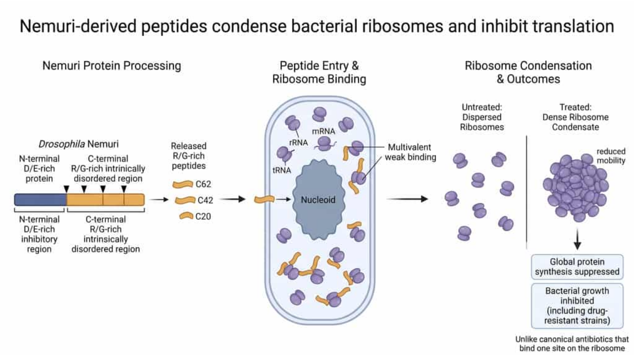
Most antibiotics that act on the ribosome bind one defined pocket. A Japanese team now reports peptides that do something different: they make the ribosome-rich regions inside a bacterium condense and lose mobility, which shuts down protein synthesis. The peptides come from Nemuri, a protein that makes fruit flies sleep and also kills bacteria.
| Measure | System | Reported result |
|---|---|---|
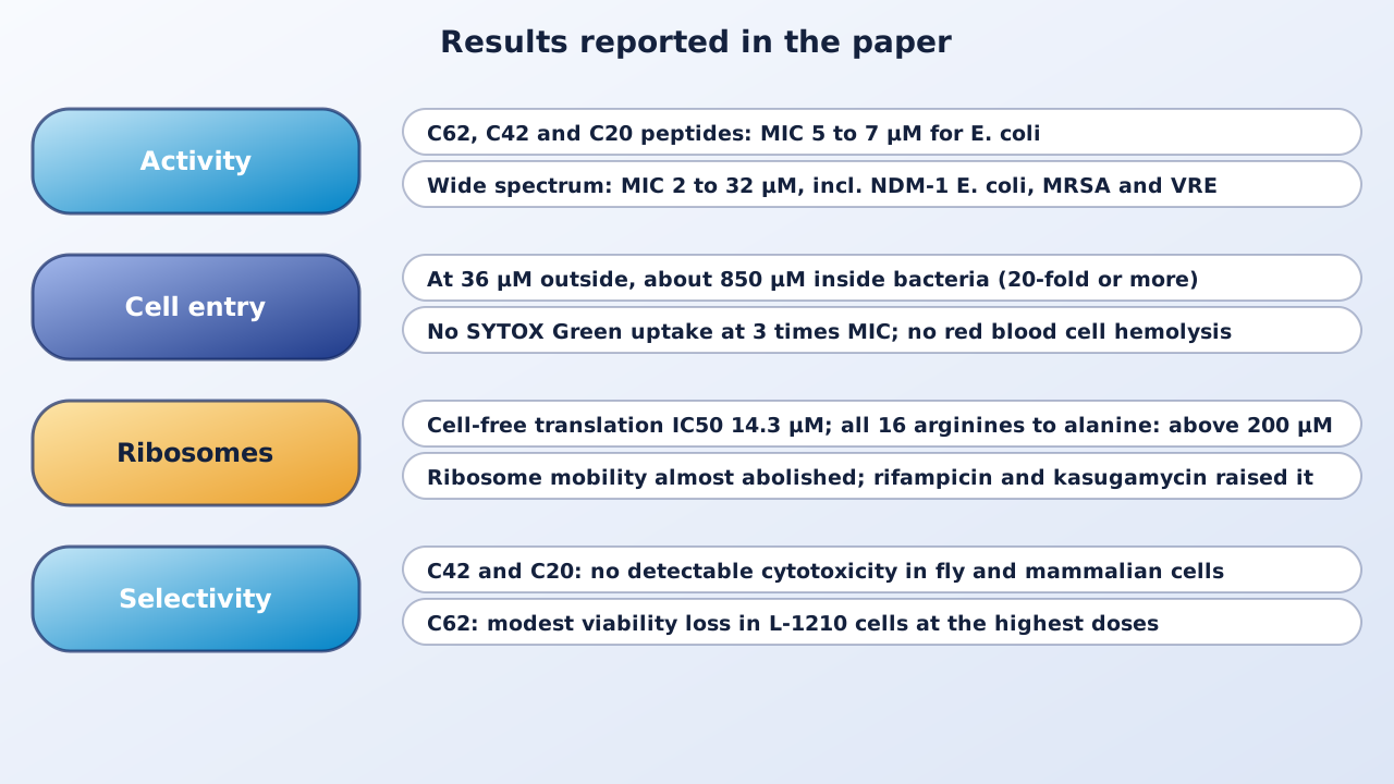
| Inhibitory concentration of C62, C42, C20 | E. coli | 5 to 7 µM |
| Range of inhibitory concentrations | Gram-negative and Gram-positive bacteria, mycobacteria, Candida | 2 to 32 µM, including NDM-1 E. coli, MRSA and VRE |
| Intracellular enrichment of C42 | E. coli, 36 µM outside | About 850 µM inside (20-fold or more) |
| Translation inhibition (IC50), C42 | Cell-free E. coli system | 14.3 µM (above 200 µM with 16 arginines replaced) |
| Proteins enriched near C42 | E. coli proximity labeling | 58 of 1,441 |
The problem and the starting point
The authors note that bacteria organize some processes in condensates and other structures, and that in Escherichia coli ribosomes sit in DNA-poor regions outside the nucleoid. That makes ribosome-rich regions a possible drug target. They had earlier found that Nemuri (Nur) kills bacteria, but how was unclear. Nur has an arginine- and glycine-rich (R/G-rich) disordered tail, a feature common in RNA-binding and condensate-forming proteins.
Cleavage releases the active peptides
Secreted full-length Nur showed no activity up to 31 µM, while short R/G-rich peptides from its tail inhibited bacteria at 9 to 10 µM and the negatively charged N-terminal half was inactive. In fly cells, Nur is cut at two sites, giving C62 and C42 fragments, and the authors also made a 20-residue piece, C20. All three inhibited E. coli at 5 to 7 µM. Replacing four RGG motifs with alanine raised the inhibitory concentration to 30 µM, replacing all 16 arginines abolished activity, and swapping arginine for lysine also reduced it (31 µM), which the authors read as a role for the arginine side chain beyond charge.
Broad activity, low toxicity to host cells
Against a panel of Gram-negative and Gram-positive bacteria, mycobacteria and Candida albicans, the peptides inhibited growth at 2 to 32 µM, including NDM-1 E. coli, multidrug-resistant Pseudomonas aeruginosa, KPC-2 Klebsiella pneumoniae, vancomycin-intermediate and methicillin-resistant Staphylococcus aureus and vancomycin-resistant Enterococcus faecalis. The concentration that kills was nearly the same as the one that inhibits. C42 and C20 showed no detectable cytotoxicity in fly S2R+ cells or in mammalian L-1210 and PANC-1 cells, and C62 caused a modest viability loss in L-1210 cells at the highest doses.
Entry without breaking the membrane
Fluorescent C42 reached the inside of E. coli within 30 minutes. At 36 µM outside, the intracellular concentration reached about 850 µM, at least 20-fold higher. Membrane stains were unchanged, SYTOX Green did not enter cells at three times the inhibitory concentration (a membrane-lytic peptide used as a control did), and red blood cells did not lyse. C42 crossed artificial lipid vesicles without disrupting them and kept its activity in a mutant lacking the SbmA transporter. In co-culture, the peptide entered bacteria at the inhibitory concentration but only weakly entered fly cells.
Binding RNA and ribosomes broadly
Proximity labeling in E. coli found 58 enriched proteins out of 1,441, including RNA helicases, rRNA-modifying enzymes and ribosomal proteins, and the peptide co-localized with a ribosomal protein marker and with RNA stains. In sucrose gradients C42 co-sedimented with 30S, 50S and 70S ribosomes in an arginine-dependent way. Cross-linking mass spectrometry showed contacts spread over the ribosome surface, with some preference for the tRNA path (uS9 and uS12), not a single site. C42 bound rRNA, mRNA and tRNA strongly and DNA only weakly.
Translation stops, and ribosomes condense
C42 lowered GFP production in E. coli without changing GFP mRNA, and reduced incorporation of two labels of new protein, azidohomoalanine and puromycin. In a cell-free system its IC50 for translation was 14.3 µM (18.4 µM for the four-alanine mutant, above 200 µM for the 16-alanine mutant). The peptides are intrinsically disordered by NMR and circular dichroism. On purified 70S ribosomes they produced visible condensates and, by cryo-EM, clustered ribosomes. In live cells ribosome-rich regions became smaller and brighter and excluded a soluble marker, and fluorescence recovery after photobleaching, which recovered within 60 seconds in untreated cells, was almost abolished. Rifampicin and kasugamycin raised ribosome mobility and chloramphenicol only modestly lowered it, so the loss of mobility is not simply a result of stopped translation.
Why it matters
The study describes a way to act on the physical organization of ribosomes rather than a single binding site, which the authors suggest could make resistance by target-site mutation less likely. It also shows how a host-defense system of an animal can use a general feature of the bacterial cytoplasm.
Keep in mind
The work is in cultured bacteria, cells and cell-free systems; no animal infection experiment is described, and there are no pharmacology, stability or dosing data in a body. How the peptides cross the membrane is unknown. The authors read the photobleaching data conservatively and say they do not show liquid-liquid phase separation or a change in the material state of the whole cytoplasm; the main physical change they detect is reduced ribosome mobility. They also note that the peptides can bind DNA weakly and inhibit transcription in simplified systems. Whether resistance arises, and whether the peptides work at useful concentrations in infection, is not tested here.
- Paper
- Nishinami S, Hasan M, Niwa T, Inui T, Yokoyama T, et al. Antimicrobial peptides derived from Nemuri induce ribosomal condensation to inhibit translation. Nature Communications. 2026;17(1):11037. Published October 29, 2026.
- Type
- Laboratory study in bacteria, cells and cell-free systems
- DOI
- 10.1038/s41467-026-78960-5
- PubMed
- PMID 42957736
- Funding
- Japan Science and Technology Agency (including CREST and PRESTO programs), Japan Society for the Promotion of Science, and the Ministry of Education, Culture, Sports, Science and Technology.
Related articles
 Daily brief · Nov 2, 2026METTL13 Methylation of eEF1A Tunes Codon Decoding, Boosts KRAS Synthesis and Is Linked to Resistance to a KRAS Inhibitor in Lung Cancer Models
 Daily brief · Nov 6, 2026Sitagliptin, a Diabetes Drug, Lowers RRM1 and RRM2 and Restores Gemcitabine Response in Models; a 22-Patient Phase 2 Trial in Pancreatic Cancer Reports a 36.4% Response Rate
 Daily brief · Nov 5, 2026Chemotherapy Raises NAD+ Recycling Through Macrophage IL-1β, and Blocking NAMPT Restores Chemotherapy Response and NK Cell Function in Mouse Tumor Models

Research focus: Molecular Oncology and Cancer Metabolism
Department of Molecular Diagnostics, Faculty of Biomedical Sciences, University of Benghazi, Libya
Weekly digest
The week's briefs and science news in one email. See the privacy policy.
Comments
No account needed. Comments are reviewed before they appear. Please be respectful and on topic.