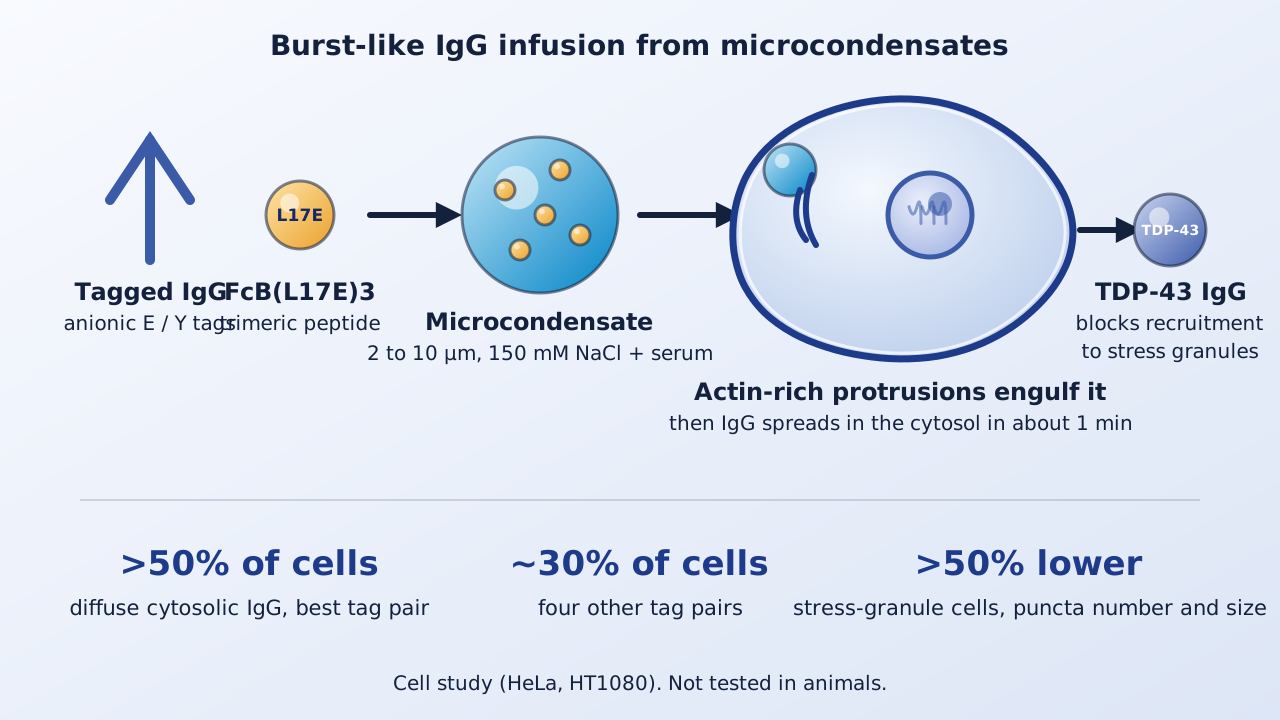
Antibodies bind targets with high specificity but rarely cross the cell membrane, which keeps most intracellular proteins out of reach. A Kyoto University group now describes a delivery method in which antibodies and a membrane-active peptide assemble into droplet-like condensates that the cell takes up and empties into the cytosol.
| Measure | System | Reported result |
|---|---|---|
| Cells with diffuse cytosolic IgG, best tag pair E20-(E4Y)2 | HeLa, 1 μM, 1 h | Over 50% |
| Other tag pairs with delivery | HeLa | About 30% (four pairs) |
| Cells with intracellular IgG signal | HeLa, E20-(E4Y)2 | 75% (nearly 50% diffuse; 25% dots only) |
| Condensate size | Holotomography | Median about 5 μm (best pair) |
| Binding of IgG to FcB(L17E)3 (KD) | SPR | 9.8 nM best; 12 to 19 nM others |
| TDP-43 stress granule recruitment | HT1080, arsenite | More than 50% reduction |
Background
Intrinsically disordered proteins such as TDP-43 are hard to inhibit with small molecules. Cell-penetrating peptides and condensate-based carriers can bring proteins into cells, but most routes end in endosomes and need hours. The group had shown that IgG carrying about 20 negative charges per molecule and a trimeric peptide FcB(L17E)3, built on the membrane-permeabilizing L17E sequence, formed microcondensates and infused IgG into cells. That earlier work used fluorescently labeled IgG.
Designing tagged antibodies
The authors fused short anionic tags of glutamic acid (E) and tyrosine (Y) to the C-terminus of the light chain of an anti-GFP IgG, away from the antigen-binding region, and later also to the heavy chain. Binding to antigen was preserved by ELISA. Light-chain tags alone formed condensates with FcB(L17E)3 in low-salt buffer but not at physiological salt. Adding tags to both chains (LHAT-IgG) produced condensates at 150 mM NaCl for all six combinations tested, with sizes of about 3 to 5 μm.
Delivery to the cytosol
With 1 μM each of IgG and peptide in serum-containing medium for 1 h, five of six LHAT-IgGs delivered IgG to the cytosol. The E20-(E4Y)2 pair worked best, with diffuse cytosolic IgG in over 50% of HeLa cells; four others reached about 30%. Direct mixing without pre-forming condensates gave no delivery. After excluding surface-bound signal, 75% of cells contained intracellular IgG and nearly 50% showed diffuse signal, while about 25% showed only dots, possibly trapped in vesicles or aggregates.
How the cell takes it up
Time-lapse and lattice light-sheet microscopy showed that condensates bound to the surface, triggered accumulation of F-actin and membrane wrapping, and were followed by cytosolic IgG release spreading in about 1 minute. Electron microscopy at 10 min showed filopodia-like protrusions reaching and penetrating condensates, with the membrane outline faded at contact sites. Cytochalasin D almost abolished diffuse cytosolic signal and methyl-β-cyclodextrin cut it to 23%, whereas chlorpromazine and nystatin had no notable effect. The authors conclude that uptake bypasses canonical endocytosis and requires cellular activity, since no delivery occurred at 4 °C.
Target binding and TDP-43
Delivered anti-GFP IgG bound its target HRas(G12V)-EGFP beneath the plasma membrane, which untagged IgG did not. In HT1080 cells expressing a cytoplasmic TDP-43 variant, pretreatment with condensates carrying an anti-TDP-43 IgG reduced, by more than 50%, the share of cells with stress granule recruitment, the number of puncta per cell and their average size after arsenite stress. An anti-pAkt IgG control produced no notable decrease.
What drives efficiency
Delivery tracked condensate number and salt stability, but not size or pairwise affinity: surface plasmon resonance gave KD of 9.8 nM for the best pair and 12 to 19 nM for the others. Condensates showed no fluorescence recovery after photobleaching, indicating a gel-like state. Tyrosine in the heavy-chain tag mattered most: replacing it with Phe or Trp lowered delivery to 36% and 32%, a Ser substitution nearly abolished it, and Asp for Glu left delivery at 52%.
Keep in mind
All results are from cultured human cell lines with model antibodies against GFP, TDP-43 and pAkt; nothing was tested in animals. The authors note that 3 to 10 μm particles raise concerns about capillary occlusion, circulation stability and immune recognition, and that local routes such as intratumoral injection might avoid them. Delivery reached about half of cells at best, and some cells kept IgG only as dots. Cell viability and effects on endogenous TDP-43 in neurons are not reported in the paper, and three authors hold a patent application on the technology.
- Paper
- Kawaguchi Y, Kiyokawa M, Yamasaki D, et al. Burst-like intracellular antibody infusion via membrane-permeabilizing peptide microcondensates. Nature Communications. 2026;17(1). Published November 16, 2026.
- Type
- Cell-culture and biophysics study
- DOI
- 10.1038/s41467-026-78720-5
- PubMed
- PMID 43039925
- Funding
- JSPS KAKENHI (JP21H04794, 23K17412, 24H00051, JP22K15250, JP24H00842); JST CREST JPMJCR18H5; JST ACT-X JPMJAX2224; JST FOREST JPMJFR2406; AMED; Integrated Research Consortium on Chemical Sciences; Kyoto University ISHIZUE 2024; JST SPRING. Three authors are inventors on a related patent application.
Related articles
 Daily brief · Nov 16, 2026A TROP2-Binding DNA Aptamer Linked to the Toxin MMAE Slowed Pancreatic Cancer Growth in Mouse Models and Added Benefit to PD-1 Blockade
 Daily brief · Nov 14, 2026Enhancer-Targeted CRISPR Activation Raised CHD8 and SCN2A Expression and Partly Rescued Mutant Phenotypes in Human Organoid and Neuron Models of Autism
 Daily brief · Nov 7, 2026Lowering MSH3 With an AAV-Delivered microRNA Cut Somatic CAG Expansion in a Huntington's Disease Mouse Model and Reduced MSH3 mRNA 48 to 94% in Macaque Striatal Neurons

Research focus: Molecular Oncology and Cancer Metabolism
Department of Molecular Diagnostics, Faculty of Biomedical Sciences, University of Benghazi, Libya
Weekly digest
The week's briefs and science news in one email. See the privacy policy.
Comments
No account needed. Comments are reviewed before they appear. Please be respectful and on topic.