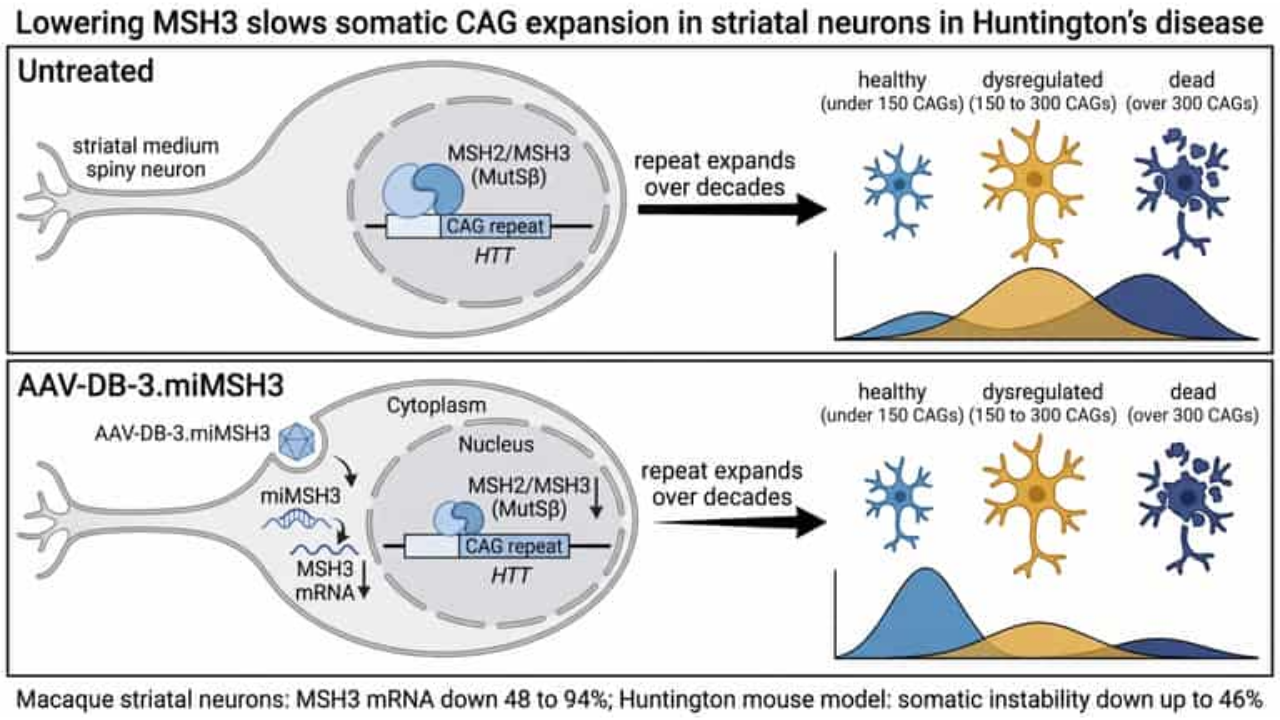
Huntington's disease is caused by an expanded CAG repeat in the huntingtin gene, and in some neurons the repeat keeps growing during life. Genetic studies point to this somatic expansion as a driver of when and how fast the disease progresses, and to a DNA repair gene, MSH3, as a way to slow it. A company-led team now reports a microRNA delivered by an AAV vector that lowers MSH3, together with a computer model that links the degree of lowering to predicted benefit.
| Measure | System | Reported result |
|---|---|---|
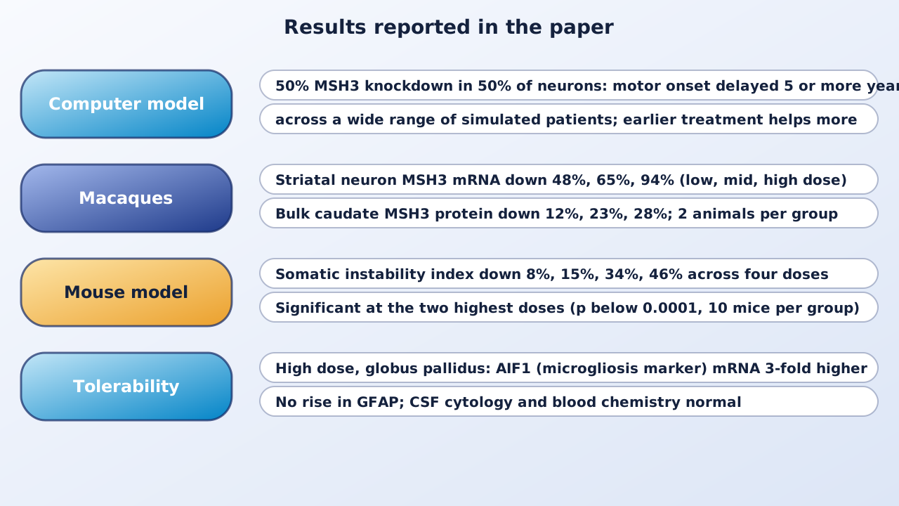
| MSH3 mRNA in striatal neurons (RNAscope) | Rhesus macaques, 2 per dose, 4 weeks | Down 48%, 65%, 94% (low, mid, high dose) |
| Bulk MSH3 protein, caudate | Same macaques | Down 12%, 23%, 28% |
| Somatic instability index | HdhQ111 mice, 10 per group, four doses | Down 8%, 15%, 34%, 46% |
| Predicted benefit of 50% lowering in 50% of neurons | Computer model | 5 or more years' delay of motor onset in a wide range of patients |
| Predicted change in cUHDRS decline, 3 years | Model, patient with 42 CAGs treated at 46 | Reduced by 53% to 126% versus natural history |
Why the repeat keeps growing
The authors describe an inherited repeat above 35 CAGs that expands further across a patient's lifetime, reaching up to 1,000 repeats in some striatal neurons in postmortem brains. They cite single-cell work showing that medium spiny neurons (MSNs) with 150 repeats or fewer keep a normal transcription profile, while those above 150 become dysregulated. MSH3 forms a complex with MSH2 that is part of mismatch repair. They cite human genetics in which a variant with about 10% lower MSH3 expression delayed onset by 1 year, and heterozygous loss-of-function mutations in MSH3 delayed average motor onset by 10.6 years. They also cite work in patient-derived neurons in which an antisense oligonucleotide that lowered MSH3 by 41% reduced somatic instability proportionally and 83% lowering roughly halted it.
A model of neuron loss and treatment benefit
The team built a simulation in which each of 3,000 MSNs carries its own CAG repeat, starting from the inherited length and expanding over the years. Neurons are called healthy under 150 CAGs, dysregulated from 150 to 300 and dead above 300. Treatment is described by two numbers: the share of neurons reached and the degree of MSH3 lowering in them. Benefit is the delay, in years, of landmarks such as motor onset (a CAP-100 equivalent score). The authors set 5 years as a meaningful threshold. In the model, 25% lowering in 25% of neurons gives 5 years of benefit only for people with short inherited repeats treated early, whereas 50% lowering in 50% of neurons gives 5 or more years for a wide range of patients. Earlier treatment helps more, and the depth of lowering matters more than reaching many neurons with a little.
The microRNA and the vector
They designed artificial microRNAs against human and macaque MSH3 mRNA and chose one, miMSH3-06, which sits in a miR-30 scaffold and works better there than in a miR-451 scaffold. It was packaged in AAV-DB-3, a capsid variant described earlier that targets MSNs and deep cortical neurons. Off-target effects were low in HEK293 cells. In human neurons carrying 49 CAG repeats, the vector lowered MSH3 mRNA by up to 52% at the highest dose, and no significant off-target changes were specific to the microRNA; mismatch repair and cancer-signaling genes did not change.
Results in macaques
Rhesus macaques received a bilateral infusion into the globus pallidus at three doses and were studied after four weeks, with two animals per group. In bulk tissue, MSH3 mRNA fell by 14%, 16% and 39% in the caudate and 9%, 16% and 24% in the putamen, and MSH3 protein fell by 12%, 23% and 28% in the caudate. Bulk measurements dilute the effect because MSNs are only 34.4% of cells in the striatum. Direct counting of MSH3 mRNA in MSNs by RNAscope showed reductions of 48%, 65% and 94% at the low, mid and high doses.
Results in a Huntington mouse model
In HdhQ111 knock-in mice, injected into the striatum at 8 weeks and examined 16 weeks later, four doses (5×10⁹ to 1.5×10¹¹ vector genomes) reduced the somatic instability index by 8%, 15%, 34% and 46%. The two highest doses were significantly different from vehicle (p below 0.0001, 10 mice per group). Striatal Msh3 mRNA fell only 13% to 27% in bulk tissue, and Msh3 protein fell by 40% at the top dose. A second microRNA, miMSH3-11, lowered mRNA by 20% and instability by 43% at one dose.
What the model predicts for the macaque data
When the macaque neuron measurements were entered into the model, all three doses exceeded the 5-year threshold, and for a subset of patients the model predicted that motor onset would never be reached. For a representative patient with 42 CAGs treated at age 46, it predicted a reduction in the decline of the composite Unified Huntington Disease Rating Scale of 53% to 126% at three years, compared with natural history. These are simulations, not trial results.
Tolerability
At the high dose, mRNA of the microgliosis marker AIF1 was 3-fold higher in the globus pallidus, which the authors call a common finding after intraparenchymal infusion. GFAP, an astrocyte marker, did not rise in any region, and cerebrospinal fluid cell counts and blood chemistry were normal. The text reports no human data.
Why it matters
The study ties a genetically supported target to a delivery method and a quantitative prediction, so that a dose can be judged by how much lowering it achieves in neurons. The authors argue the model applies to any approach that lowers somatic instability, whether antisense, RNA interference, gene editing or small molecules, and that it could be adapted to other repeat disorders.
Keep in mind
The macaque result rests on two animals per dose over four weeks, and the clinical benefit is a prediction from a model built on a small number of donors, not a measured outcome. The mouse model shows reduced instability but lacks robust functional features, so neuron survival and behavior were not tested. Cortical instability was not modeled. Delivery here is by direct infusion into the brain. Most authors are founders or employees of Latus Bio, the company developing the therapy, and the work was funded in part by it. No clinical results are reported.
- Paper
- Simpson BP, Ranum PT, Leib DE, et al. Computational modeling and preclinical evaluation support targeting somatic instability for Huntington’s disease treatment. Nature Communications. 2026;17(1):11036. Published October 28, 2026.
- Type
- Computer model with macaque and mouse studies
- DOI
- 10.1038/s41467-026-78769-2
- PubMed
- PMID 42951910
- Funding
- Latus Bio, the CHOP Research Institute, and the Hereditary Disease Foundation Transformative Research Award (115873). Several authors are founders or employees of Latus Bio.
Related articles
 Daily brief · Nov 1, 2026Engineered Retrotransposons Called Gene Writers Insert Gene-Length DNA From an RNA Template, Including to Make CAR-T Cells In Vivo in Mice
 Daily brief · Oct 31, 2026First-in-Human AAV-CRISPR Therapy for Duchenne Muscular Dystrophy Shows Editing in Muscle but a Fatal Immune Reaction at the Higher Dose
 Daily brief · Oct 21, 2026A Compact RNA Switch Lets a Small-Molecule Drug Turn Gene Therapy On and Off in Mice

Research focus: Molecular Oncology and Cancer Metabolism
Department of Molecular Diagnostics, Faculty of Biomedical Sciences, University of Benghazi, Libya
Weekly digest
The week's briefs and science news in one email. See the privacy policy.
Comments
No account needed. Comments are reviewed before they appear. Please be respectful and on topic.