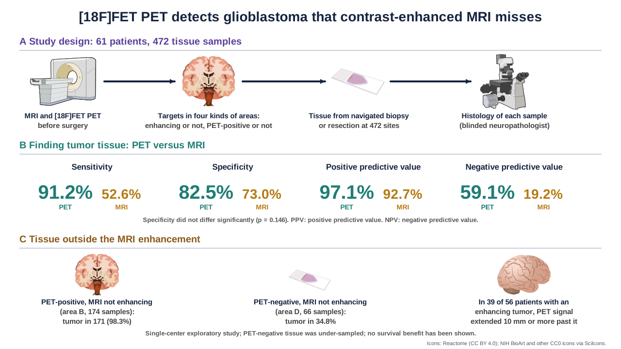
Surgeons and radiation oncologists plan around the part of a glioblastoma that lights up on contrast-enhanced MRI, but the tumor often extends beyond it. A team at LMU Munich and partner centers now reports that PET with the amino acid tracer O-(2-[18F]fluoroethyl)-L-tyrosine ([18F]FET) found far more tumor tissue than MRI in 472 tissue samples, each checked under the microscope.
| Measure (472 samples, 61 patients) | [18F]FET PET | Contrast-enhanced MRI |
|---|---|---|
| Sensitivity | 91.2% | 52.6% |
| Specificity | 82.5% | 73.0% (p = 0.146) |
| Positive predictive value | 97.1% | 92.7% |
| Negative predictive value | 59.1% | 19.2% |
| Tumor in PET-positive, non-enhancing tissue | 171 of 174 (98.3%) | Not applicable |
Why the question matters
Glioblastoma, IDH wildtype, is the most common malignant brain tumor in adults, and the extent of resection is one of its strongest prognostic factors. MRI shows a tumor when the blood-brain barrier is disrupted and a contrast agent leaks in, so infiltrating tumor without enhancement can be missed. T2/FLAIR MRI shows more, but changes such as edema and scarring also light up. The authors note that earlier studies reported larger tumor volumes on amino acid PET, but histology-verified data with many sampling sites were scarce.
How the study was done
From October 2019 to July 2025, 147 patients with suspected glioma were enrolled prospectively at one center. This analysis includes the 61 whose tumor was confirmed as IDH-wildtype glioblastoma: 45 newly diagnosed and 16 recurrent. All had MRI and [18F]FET PET before surgery. Targets were chosen on the coregistered scans in four kinds of areas: enhancing and PET-positive (area A), non-enhancing and PET-positive (area B), enhancing and PET-negative (area C), and neither (area D). PET-positive meant a tumor-to-background ratio of 1.6 or more. Samples came from stereotactic biopsy in 26 patients and from resection in 35, were tracked by navigation coordinates, and were graded by a neuropathologist blinded to clinical data as no tumor, infiltration zone or solid tumor. There were 472 samples in all: 210 from area A, 174 from B, 22 from C and 66 from D.
What PET found that MRI did not
PET uptake was present in all 61 patients, while contrast enhancement was present in 56. Across all samples, PET had a sensitivity of 91.2% against 52.6% for MRI (p below 0.001), with similar specificity (82.5% against 73.0%, p = 0.146). Positive predictive value was 97.1% for PET and 92.7% for MRI, and negative predictive value was 59.1% against 19.2%. In area B, where MRI showed no enhancement, 171 of 174 samples held tumor (98.3%). In area D, neither modality was positive and 34.8% of samples still held tumor. In 39 of 56 patients with an enhancing tumor (69.6%), PET signal extended at least 10 mm past the enhancing margin. In the five patients with no enhancement at all, all 30 area B samples held tumor.
Density and T2/FLAIR
In tumor-containing samples, PET detected 82.3% of those in the infiltration zone against 43.2% for MRI, and 99.1% of solid tumor against 60.8%. The PET signal rose with tumor burden (Spearman correlation 0.58). T2/FLAIR was less specific than PET. Among samples that were non-enhancing and PET-negative but T2/FLAIR-positive, only 37.0% (17 of 46) held tumor.
What the authors suggest
They propose that PET could guide supramarginal resection and radiotherapy target definition beyond the enhancing margin, and that PET-based response criteria could be used in follow-up, and they call for prospective trials. They note that a recent trial (GLIAA) found no outcome advantage for PET-based over MRI-based target delineation in re-irradiation of recurrent glioblastoma, and that this finding should be read cautiously.
Keep in mind
This is a single-center exploratory study. Sampling was targeted and opportunistic, with several samples per patient, and PET-negative tissue was under-sampled because extra biopsies carry risk, so the negative predictive value of 59.1% limits conclusions about tissue without PET signal. Recurrent disease was a small subgroup with no area D samples, and brain shift during surgery could have affected the match between images and samples. The study shows that PET is more sensitive for finding tumor tissue; it does not show that using PET in surgery or radiotherapy improves survival.
- Paper
- Stürzl R, Kirchleitner SV, Lorenz J, Schmidbauer KJ, Stöcklein V, Quach S, et al. [18F]FET PET enables histopathology-validated detection of non-contrast-enhancing glioblastoma. Nature Communications. 2026;17(1). Published November 6, 2026.
- Type
- Prospective single-center exploratory diagnostic study
- DOI
- 10.1038/s41467-026-78980-1
- PubMed
- PMID 42995172
- Funding
- Deutsche Forschungsgemeinschaft (DFG) research unit FOR-2858 (project numbers 421887978, 422188432, 403161218 and 422179811) and the Thomas Kirch Stiftung; the funders had no role in the study. Several authors report honoraria, advisory roles or research funding from companies, including Telix Pharmaceuticals and Novocure; the full statement is in the paper.
Related articles
 Feature · Oct 19, 2026Why a Glutaminolysis Signature Falls With PTEN Loss: Passenger Co-Deletion, Not Metabolic Regulation
 Daily brief · Nov 15, 2026In Enzalutamide-Resistant Prostate Cancer Cells, m6A Marking of ARv7 mRNA Raises Its Translation, Lactate Feeds the Loop, and a METTL3 Inhibitor Plus Enzalutamide Slowed Xenograft Growth
 Daily brief · Nov 11, 2026Higher Blood Levels of Immune Proteins, Including IL15 and FLT3LG, Measured Years Before Diagnosis Were Linked to Lower Prostate Cancer Risk, and Genetics Pointed to PARP1, SPINT2 and NME3

Research focus: Molecular Oncology and Cancer Metabolism
Department of Molecular Diagnostics, Faculty of Biomedical Sciences, University of Benghazi, Libya
Weekly digest
The week's briefs and science news in one email. See the privacy policy.
Comments
No account needed. Comments are reviewed before they appear. Please be respectful and on topic.