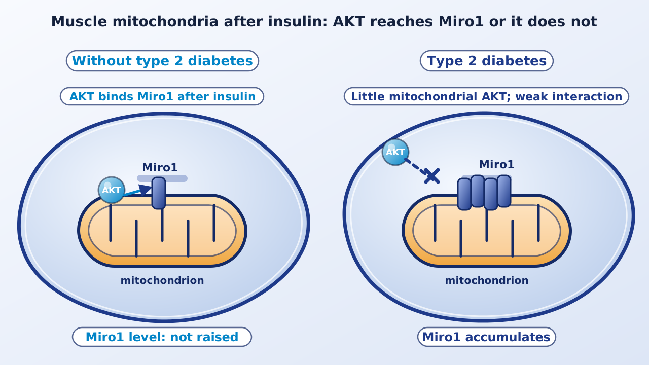
Skeletal muscle takes up most of the glucose the body clears after a meal, so muscle that responds poorly to insulin is a central defect in type 2 diabetes. A group at the Pennington Biomedical Research Center now reports that a mitochondrial protein called Miro1 accumulates in the muscle of patients with type 2 diabetes, and that its removal in mice improves how muscle handles glucose.
Background. Type 2 diabetes affects more than 500 million people worldwide. Skeletal muscle accounts for about 85% of whole-body glucose disposal at rest, and obesity is present in more than 90% of patients. Mitochondria in muscle fuse, divide and are recycled, and patients with type 2 diabetes show mitochondrial defects. Miro1 (mitochondrial Rho GTPase 1) sits on the outer mitochondrial membrane, can anchor the fission protein DRP1, and was known mainly for moving mitochondria along nerve cell axons. Its role in muscle and in diabetes was unknown. The brief on mitochondria and ferroptosis covers a different mitochondrial question.
Miro1 accumulates only in type 2 diabetes. In a cross-sectional study, adults were assigned to healthy weight, obesity without diabetes, or obesity with type 2 diabetes. Each had a three-day inpatient stay with a fasting muscle biopsy and a hyperinsulinemic-euglycemic clamp, the reference test of insulin sensitivity. Miro1 protein was raised only in the patients with type 2 diabetes, and it correlated modestly and negatively with insulin sensitivity, a relation the authors say is largely driven by that group. The same accumulation appeared in mice fed a high-fat, high-fructose, high-cholesterol diet and in two genetic mouse models of diabetes, so the authors conclude it tracks insulin resistance and not obesity alone.
Why it builds up. In muscle cells, insulin lowered Miro1 and raised its co-localization with AKT to about 60%. In the patients, insulin-stimulated AKT phosphorylation at threonine 308 in isolated mitochondria was almost absent in type 2 diabetes compared with obesity alone. When the AKT inhibitor MK-2206 was added with insulin, Miro1 accumulated quickly. The authors conclude that inadequate insulin-driven AKT translocation to mitochondria, and so inadequate AKT-Miro1 interaction, explains the build-up.
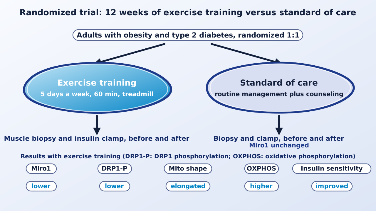
Exercise training lowers it. The DYNAMMO-T2D trial (NCT02977442) randomized adults aged 18 to 60 with obesity and type 2 diabetes, 1:1, to 12 weeks of supervised treadmill training or standard care. Training was five days a week for an hour at 60% to 85% of maximal heart rate. Insulin sensitivity improved with training and not with standard care. Miro1 fell, as did DRP1 phosphorylation and the DRP1 recruiters Mid49 and Mid51; muscle mitochondria became less fragmented and more elongated; and maximal oxidative phosphorylation rose. Markers of mitochondrial biogenesis and fusion, including PGC1α, MFN1, MFN2 and OPA1, did not increase, which the authors read as remodeling of the network rather than making new mitochondria.
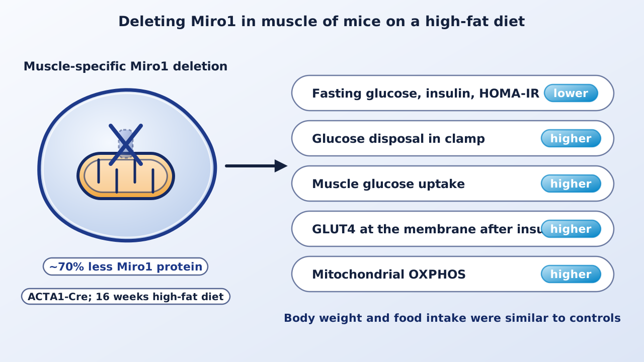
Deleting Miro1 in muscle. Because exercise acts on many tissues, the authors deleted Miro1 only in skeletal muscle of male mice (about 70% less protein) and fed them a high-fat diet for 16 weeks. Body weight and food intake matched controls, while fasting glucose, fasting insulin and HOMA-IR were lower, insulin tolerance and glucose disposal in the clamp were better, muscle glucose uptake was higher, AKT and AS160 signaling and GLUT4 at the membrane were greater, and mitochondrial respiration was higher. Knocking down Miro1 in cultured C2C12 muscle cells increased glucose uptake and respiration as well.
Why it matters. The work places a mitochondrial protein on the path between insulin signaling and glucose uptake, and it gives a molecular reading of one benefit of exercise: it clears a protein that holds muscle back. The authors propose Miro1 as a possible therapeutic target. They also note the opposite pattern in the pancreas, where Miro1 is reported to be depleted in type 2 diabetes, so tissue-specific effects would have to be considered.
Keep in mind. The human findings are associations and a single randomized trial of exercise, which cannot show that lower Miro1 alone explains the gain in insulin sensitivity; exercise changes many things at once. The trial was open-label. The causal evidence comes from mice and cultured cells, and the deletion work used male mice. The authors state that prospective human studies are needed to see whether Miro1 builds up as people move from prediabetes to type 2 diabetes. Miro1 is not an established drug target, and no treatment based on it exists.
- Paper
- Heintz EC, Dantas WS, Taylor AL, et al. Miro1 mediates skeletal muscle insulin resistance in type 2 diabetes. Nature Communications. 2026;17(1). Published online October 19, 2026. Open access (PMC13680474). ClinicalTrials.gov NCT02977442.
- Type
- Human cross-sectional study, randomized trial and mouse experiments
- DOI
- 10.1038/s41467-026-77278-6
- PubMed
- PMID 42897135
Related articles
 Daily brief · Oct 19, 2026A New Class of Mitochondria-Targeted Compounds Induces Ferroptosis and Suppresses Tumors in Cancer Models
 Explainer · Oct 26, 2026How a Personalized mRNA Cancer Vaccine Works, and What the Melanoma Trials Show So Far
 Science news · Oct 24, 2026First Prime Editing Treatment Given Directly in the Body Enters a Wilson Disease Trial

Research focus: Molecular Oncology and Cancer Metabolism
Department of Molecular Diagnostics, Faculty of Biomedical Sciences, University of Benghazi, Libya
Weekly digest
The week's briefs and science news in one email. See the privacy policy.
Comments
No account needed. Comments are reviewed before they appear. Please be respectful and on topic.