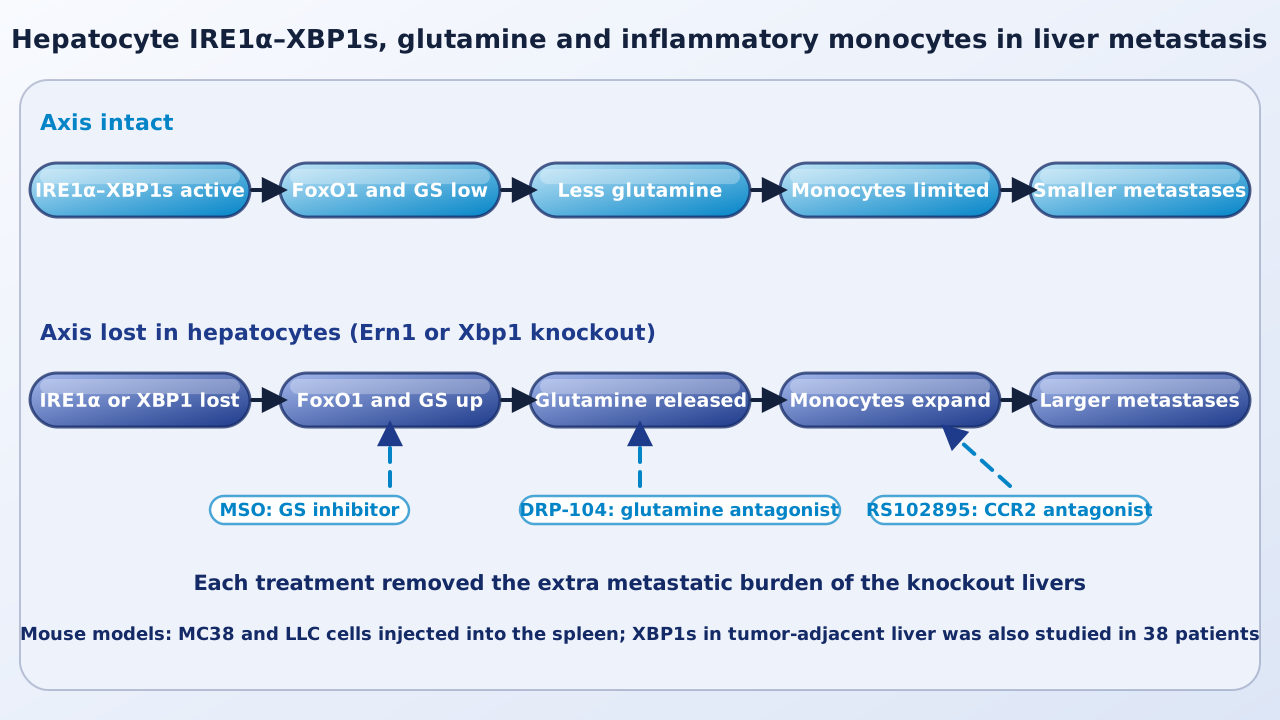
Drugs that block IRE1α, a sensor of stress in the endoplasmic reticulum, shrink many mouse tumors. A group at the QIMR Berghofer Medical Research Institute, the University of Macau and Sun Yat-sen University now reports that the same drug fails against metastases in the liver, and that the reason lies in the healthy liver cells around the tumor, not in the cancer cells.
| Experiment | System | Reported result |
|---|---|---|
| IRE1α inhibitor MKC8866 | MC38 and LLC tumors, 6 mice per group | Reduced subcutaneous growth; no reduction of MC38 liver metastases |
| Hepatocyte deletion of Ern1 or Xbp1 | MC38 and LLC liver metastasis, 6 mice per group | Larger liver metastases than littermate controls |
| Single-cell RNA sequencing | 7,515 liver myeloid cells at day 7 | Monocyte cluster enlarged in knockouts |
| Glutamine release by hepatocytes | Primary cells, 0.05 mM glutamine medium | 30% higher in Ern1 knockout supernatant |
| Colorectal cancer liver metastases | 38 patients, 19 per XBP1s group | Higher XBP1s in adjacent liver: less recurrence or better survival |
Why the question matters
IRE1α and its downstream transcription factor XBP1s promote tumors through several cell types, and an IRE1α inhibitor, ORIN1001 (MKC8866), has been tested in people. The authors note that a phase 1/2 trial in advanced solid tumors (NCT03950570) has not given encouraging results. The registry lists the study as completed in January 2023 with 58 participants enrolled and no results posted. The liver is a common site of metastasis; the authors cite liver involvement in up to 80% of patients with pancreatic cancer and 50% of those with colorectal cancer. Most work on IRE1α had looked at the tumor itself, not at the liver tissue that receives the metastasis.
An IRE1α inhibitor did not reach liver metastases
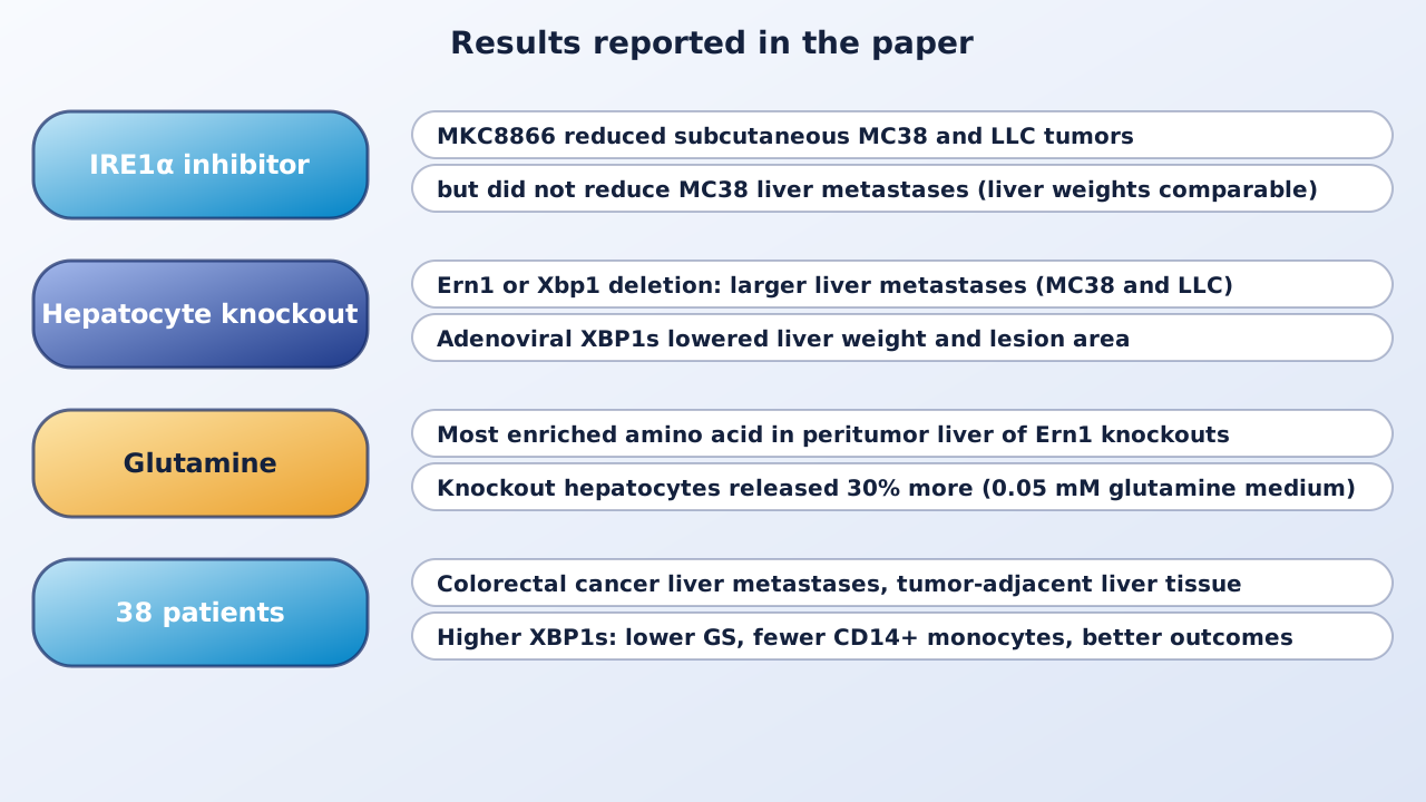
In mice, MKC8866 reduced the growth of subcutaneous tumors formed by MC38 colon and LLC lung cancer cells. When the same cells were injected into the spleen to seed the liver, the inhibitor did not lower the liver weight of MC38-bearing mice, and in LLC-bearing mice liver weight was slightly higher. In a 4T1 breast cancer model with spontaneous metastasis, MKC8866 reduced lung metastases but gave no benefit in the liver. The authors concluded that IRE1α–XBP1s signaling in the liver might act against tumors in a way that opposes its tumor-intrinsic effect.
Deleting the axis in hepatocytes enlarged metastases
The team deleted Ern1 (the gene for IRE1α) or Xbp1 specifically in mouse hepatocytes. With six mice per group, both knockouts had larger liver metastases than littermate controls, for MC38 and LLC cells. Delivering XBP1s to hepatocytes with an adenovirus reduced liver weight and the liver area covered by lesions. The first days were similar: micrometastasis numbers did not differ at day 2, but lesions were larger in the knockouts by day 7, with more Ki67-positive cells and no difference in apoptosis. The authors therefore describe an effect on outgrowth, not on first colonization.
Glutamine and CCR2-positive monocytes
Metabolomics of peritumor liver at day 7 showed glutamine as the most enriched amino acid in Ern1 knockout livers. The glutamine antagonist DRP-104 removed the extra metastatic burden, and MC38 cells lacking the glutamine transporter SLC38A2 formed fewer liver metastases. Immune profiling pointed to a specific cell type. Natural killer and CD8 T cell depletion did not change the difference between genotypes, but an antibody against Ly6C, which depletes circulating monocytes, did. Single-cell RNA sequencing of 7,515 liver myeloid cells (3,715 from controls, 3,800 from knockouts) showed an enlarged monocyte cluster rich in Ccr2 in knockouts, and the CCR2 antagonist RS102895 abolished the difference between genotypes. Glutamine given at day 6 increased CCR2-positive monocytes and sped metastatic progression in normal mice, while at day 1 it did nothing and in Ccr2-deficient mice it did not promote metastasis.
How hepatocytes control glutamine
Primary hepatocytes from knockouts released more glutamine: in medium with 0.05 mM glutamine, the supernatant of Ern1 knockout cells held 30% more than that of control cells, and Xbp1 knockout cells behaved the same way. Medium from knockout hepatocytes expanded bone marrow monocytes in culture, and L-asparaginase, which breaks down glutamine, reduced that effect. In the liver, hepatocytes next to metastases in knockouts expressed glutamine synthetase (GS) and FoxO1, a transcription factor that can switch on the GS gene. A FoxO1 inhibitor lowered GS and glutamine release in cultured knockout hepatocytes, and the GS inhibitor methionine sulfoximine removed the difference in metastatic burden and in monocyte numbers. A form of XBP1s lacking its DNA-binding domain still lowered glutamine release, which the authors read as a transcription-independent effect.
Patients with colorectal cancer liver metastases
The authors examined liver tissue from 38 patients who had neoadjuvant chemotherapy and then surgery for colorectal cancer liver metastases at Sun Yat-sen University Cancer Center between 2012 and 2016. In tumor-adjacent hepatocytes, higher XBP1s went with lower GS and fewer CD14-positive monocytes at the invasive margin (19 patients per group). Patients with higher XBP1s in adjacent liver, or a higher liver-to-tumor XBP1s ratio, had less recurrence or better survival by Kaplan-Meier analysis; the authors report the highest liver-to-tumor ratio had the most favorable outcome.
Why it matters
The study adds a metabolic mechanism for why a drug that works on the primary tumor can fail in a metastatic organ: healthy tissue around the tumor can change the nutrient supply to immune cells. It also shows that glutamine acts here through monocytes, not only through cancer cells. The authors conclude that IRE1α antagonism will likely need careful design and combinations, and that peritumor hepatocytes are a possible target.
Keep in mind
Nearly all the causal experiments are in mice, with two injected cancer cell lines (MC38 and LLC) and complete, hepatocyte-specific knockouts, which differ from a drug given to a person. The patient work is a correlation in 38 people from one center and does not show that XBP1s protects them. The authors state that clinical data on anti-metastatic efficacy are not yet available. Whether glutamine or GS inhibitors would work in people with liver metastases is untested here, and IRE1α–XBP1s also supports normal liver function, so blocking it broadly has other consequences.
- Paper
- Zhang H, Liang P, Yang W, et al. Hepatocyte IRE1α–XBP1s axis prevents pro-metastatic niche formation through metabolic rewiring of inflammatory monocytes. Nature Communications. 2026;17(1):10961. Published November 3, 2026.
- Type
- Mouse models with patient tissue analysis
- DOI
- 10.1038/s41467-026-78812-2
- PubMed
- PMID 42977746
- Funding
- Guangdong Provincial Science Fund for Distinguished Young Scholars (2021B1515020007), Macau Science and Technology Development Fund (0071/2023/RIA2 and 0059/2024/ITP2), and the General Program of Natural Science Foundation of Guangdong (2024A1515013027).
Related articles
 Daily brief · Nov 3, 2026A Network Model of the Tipping Point in Colon Tumor Development Points to USP7, and a USP7 Inhibitor Shifts Cancer Organoids Toward a Normal-Like State
 Feature · Oct 19, 2026Glutamine Plus Chemotherapy for Advanced Pancreatic Cancer: A Small Phase 1 Trial Finds the Combination Feasible
 Explainer · Oct 19, 2026Glutamine Addiction: Why Some Cancer Cells Depend on a Nonessential Amino Acid

Research focus: Molecular Oncology and Cancer Metabolism
Department of Molecular Diagnostics, Faculty of Biomedical Sciences, University of Benghazi, Libya
Weekly digest
The week's briefs and science news in one email. See the privacy policy.
Comments
No account needed. Comments are reviewed before they appear. Please be respectful and on topic.